
Welcome to my article on the fascinating topic of the external anatomy of the anterior heart. In this piece, we will delve into the intricate details of the structure and function of the anterior heart, shedding light on its vital role in our cardiovascular system.
As we explore the external anatomy of the anterior heart, we will uncover the unique features that make it such a remarkable organ. From its size and shape to the placement of its major blood vessels, we will examine how these elements work together to ensure the efficient flow of blood throughout our bodies.
What is the Anterior Heart?
The anterior heart refers to the front part of the heart, specifically the structures that can be observed and studied from an external perspective. It plays a crucial role in the functioning of the cardiovascular system, responsible for pumping oxygenated blood to all parts of the body. Understanding the external anatomy of the anterior heart is essential for correctly identifying and labeling its different components. In this section, I will explore the key features of the anterior heart, highlighting its unique structures and their functions.
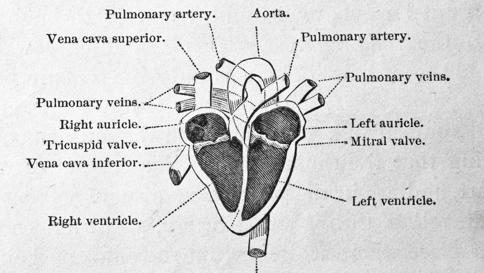
The external anatomy of the anterior heart includes several distinct features that contribute to its efficient functioning. These features can be classified into four main components: the chambers, the valves, the major vessels, and the coronary arteries. Let’s take a closer look at each of these components:
- Chambers:
- The anterior heart consists of four chambers: two atria and two ventricles.
- The atria, located at the top, receive blood from the body and lungs.
- The ventricles, situated at the bottom, pump blood out of the heart to various parts of the body.
- Valves:
- The anterior heart has four valves that ensure the unidirectional flow of blood.
- The atrioventricular (AV) valves, including the mitral (bicuspid) and tricuspid valves, separate the atria from the ventricles.
- The semilunar valves, including the aortic and pulmonary valves, control the blood flow out of the heart.
- Major Vessels:
- The anterior heart is connected to major blood vessels.
- The superior and inferior vena cava bring deoxygenated blood from the body into the right atrium.
- The pulmonary veins carry oxygenated blood from the lungs into the left atrium.
- The aorta is the largest artery that carries oxygenated blood from the left ventricle to the rest of the body.
- Coronary Arteries:
- The anterior heart has its own blood supply through the coronary arteries.
- The left coronary artery and right coronary artery branch off from the aorta to provide oxygenated blood to the heart muscle.
Correctly Label The Following External Anatomy Of The Anterior Heart.
When it comes to studying the external anatomy of the anterior heart, understanding its size and shape is crucial for accurately identifying and labeling its various components. The size and shape of the anterior heart can vary slightly between individuals, but there are some general characteristics that can help guide us in the process of correctly labeling its external anatomy.
1. Size: The size of the anterior heart can be described as approximately the size of a closed fist. It typically measures about 12 centimeters in length and 8 centimeters in width. However, it’s important to note that the size of the heart can vary depending on factors such as age, gender, and overall health.
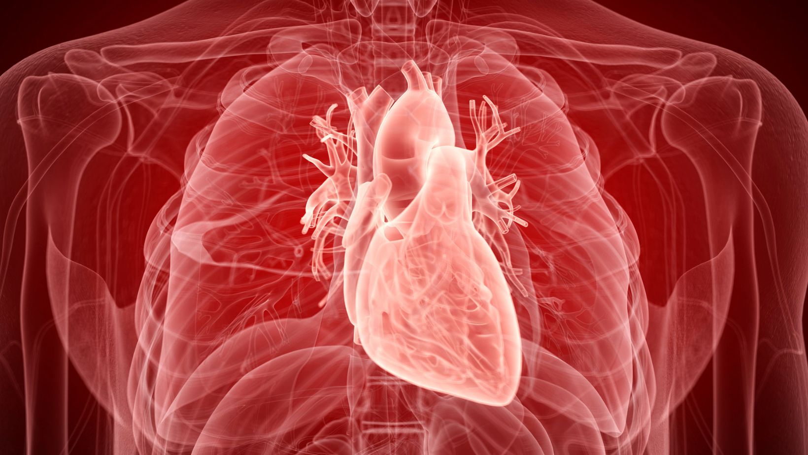
2. Shape: The shape of the anterior heart can be likened to that of a cone. It has a pointed apex at the bottom and a broader base at the top. The apex of the heart is directed downward and to the left, while the base is located at the top and is formed by the atria. The overall shape of the anterior heart allows it to fit snugly within the protective confines of the ribcage.
3. Ventricular Mass: The anterior heart is primarily composed of the left and right ventricles, which are responsible for pumping oxygen-rich blood to the rest of the body. The left ventricle, in particular, is the largest and strongest of the four chambers, as it needs to exert enough force to propel blood throughout the entire circulatory system.
4. External Features: When examining the external anatomy of the anterior heart, you’ll notice some key features that aid in its identification. These include the coronary sulcus (also known as the atrioventricular groove), which separates the atria from the ventricles, and the interventricular sulci, which divide the ventricles into distinct regions. Additionally, the anterior heart is covered by a protective layer called the pericardium, which provides support and helps reduce friction.
Understanding the size and shape of the anterior heart is crucial for correctly labeling its external anatomy. With this knowledge, we can efficiently navigate through the chambers, valves, major vessels, and coronary arteries, ensuring accuracy in our understanding of this vital organ’s structure. So now that we have a solid foundation of the anterior heart’s proportions, let’s delve further into its external components and their functions, allowing us to paint a comprehensive picture of this remarkable organ.